As noted in this prior post, it has been particularly exciting to learn from A/Prof Caroline Hauxwell’s (Faculty of Science, School of Biology & Environmental Science.) extensive knowledge of the site and its soil biology. (We had also engaged in fruitful discussions with here prior about the setup and the direction of the regeneration project).
Dr. Hauxwell has been using adjacent areas at SERF and part of the artwork/revegetation site for her research experiments into beneficial soil fungi – which have an end outcome to reduce pasture dieback that is caused by mealy bugs. (The bug in question is the paspalum mealybug, Heliococcus summervillei – and her work is of particular relevance to the pasture industry given that these invasive bugs devour Buffalo grass which is a key, commercial pasture grass, (and interestingly a notable environmental weed in conservation contexts). To do this work Caroline and her research team have become experts in isolating fungi from soils of forests and pastures.
Immediately before, and following the burn of the SERF wet gulley/artwork site A/Prof Hauxwell had initiated a periodic soil sampling regime to learn how the fungal composition of the site might change according to the burn – something which also seemed very relevant to us given we needed proxy ways to determine the improvement of soil and plant health as the artwork process evolves.
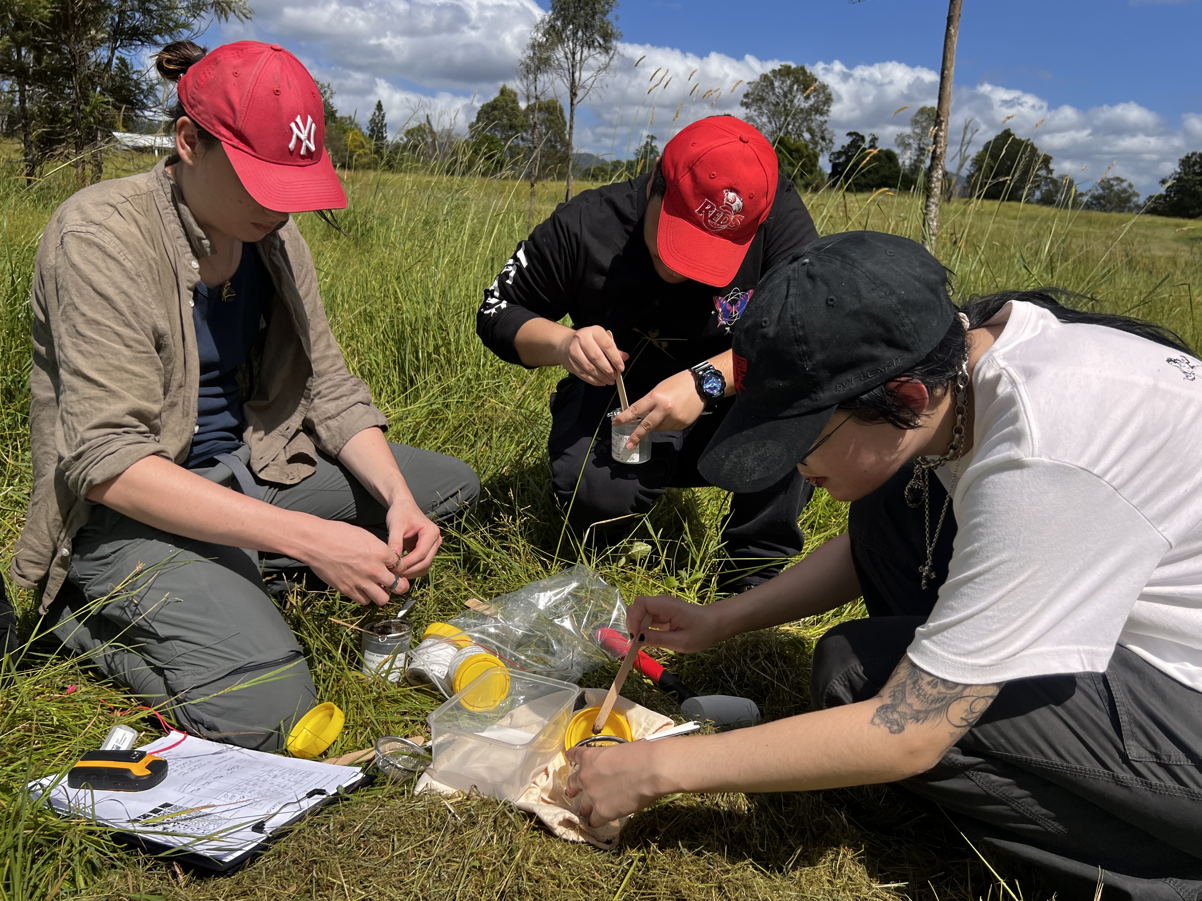
So on 11/3/24 Dr. Eleanor Velasquez and I joined the soil sampling process at SERF – to understand and observe more about the scientific methods underway.
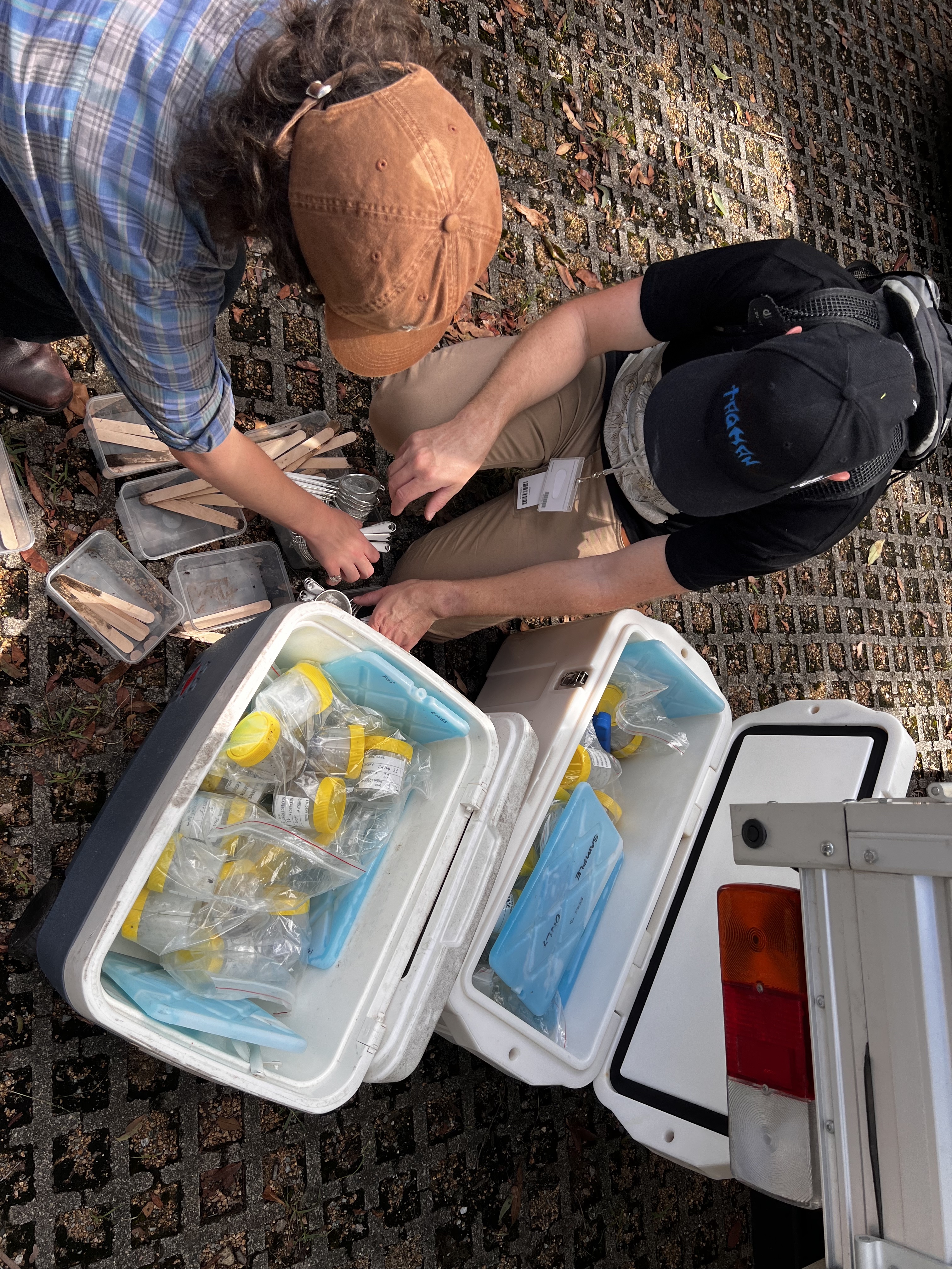
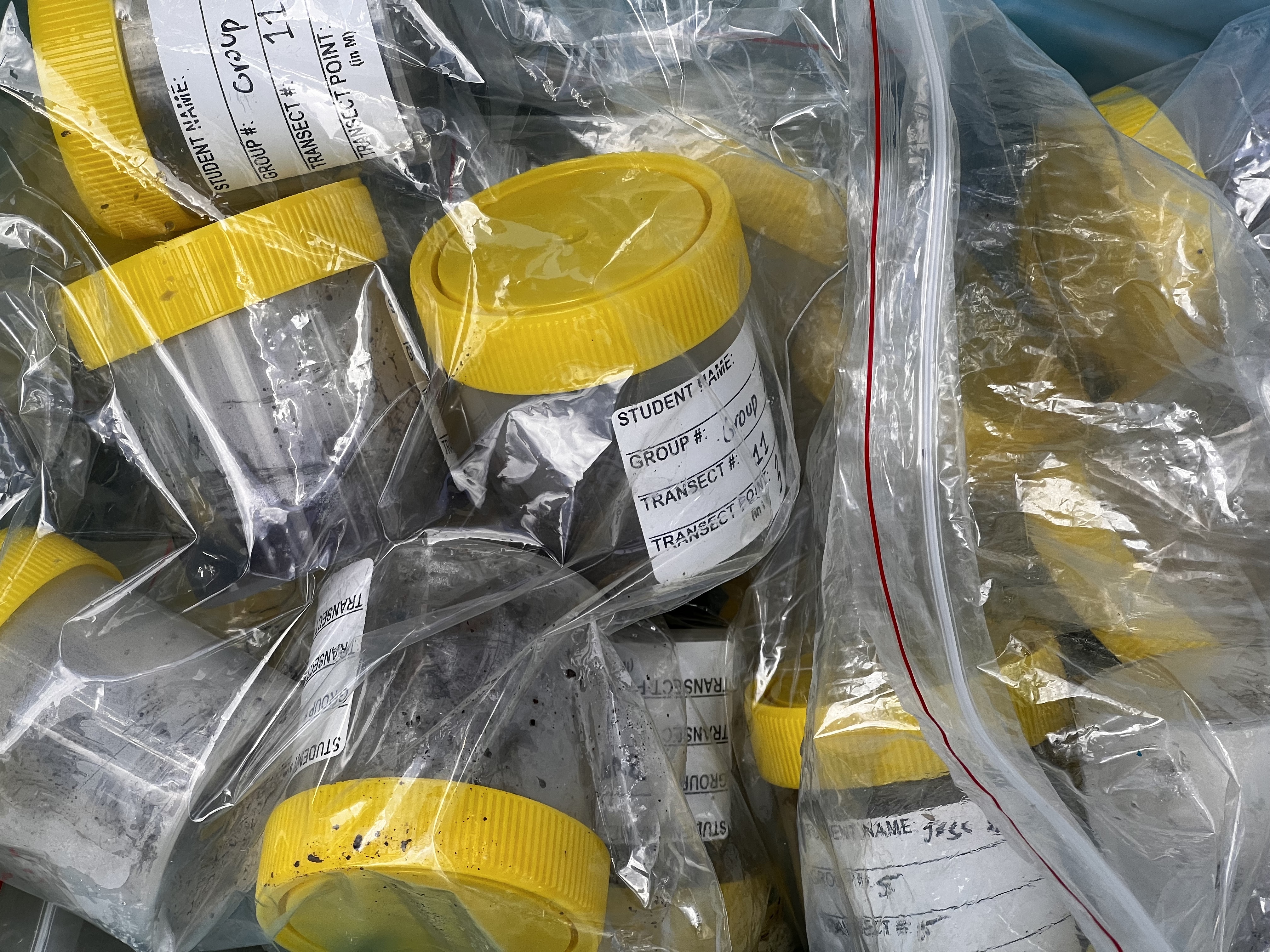
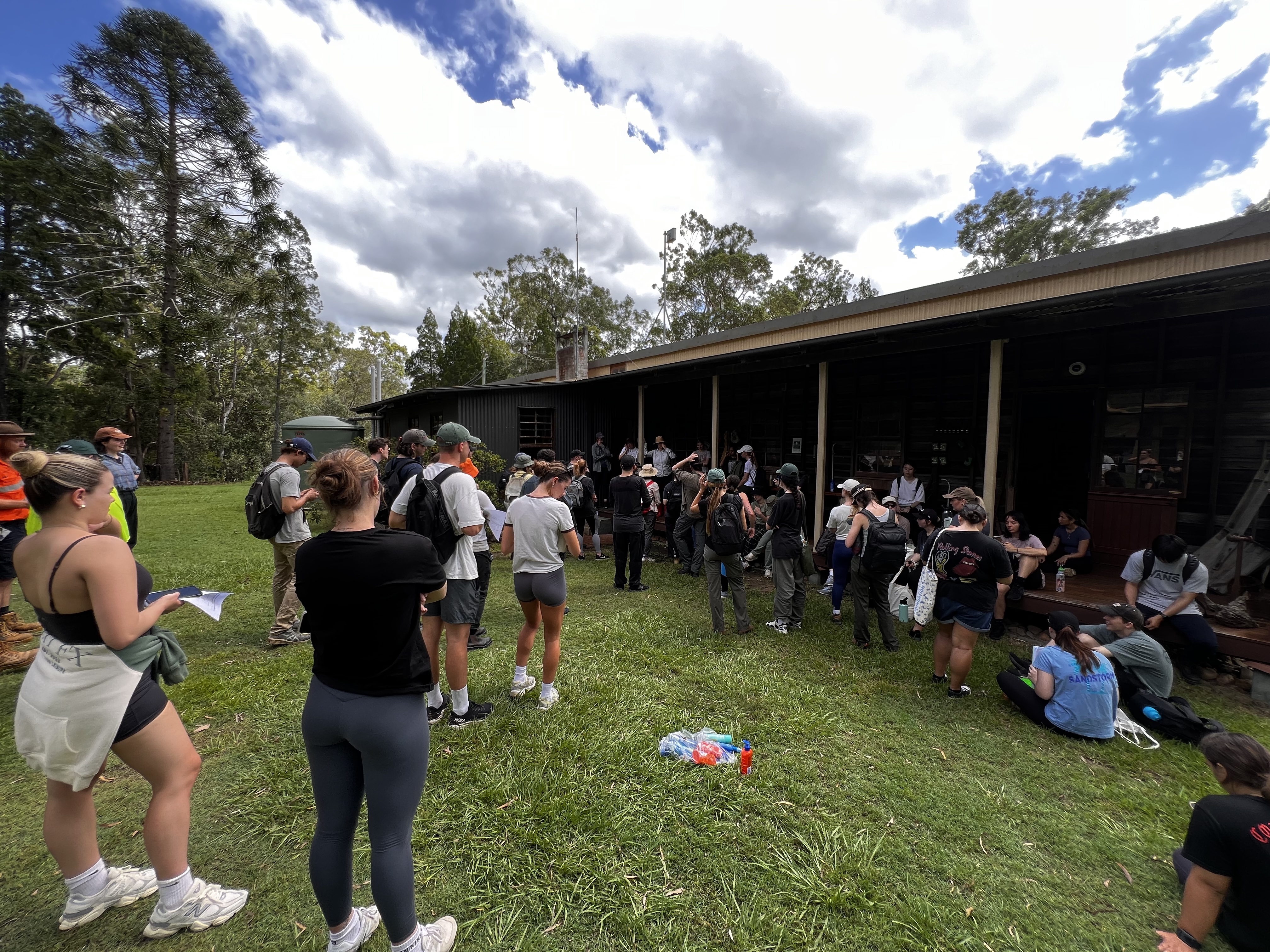
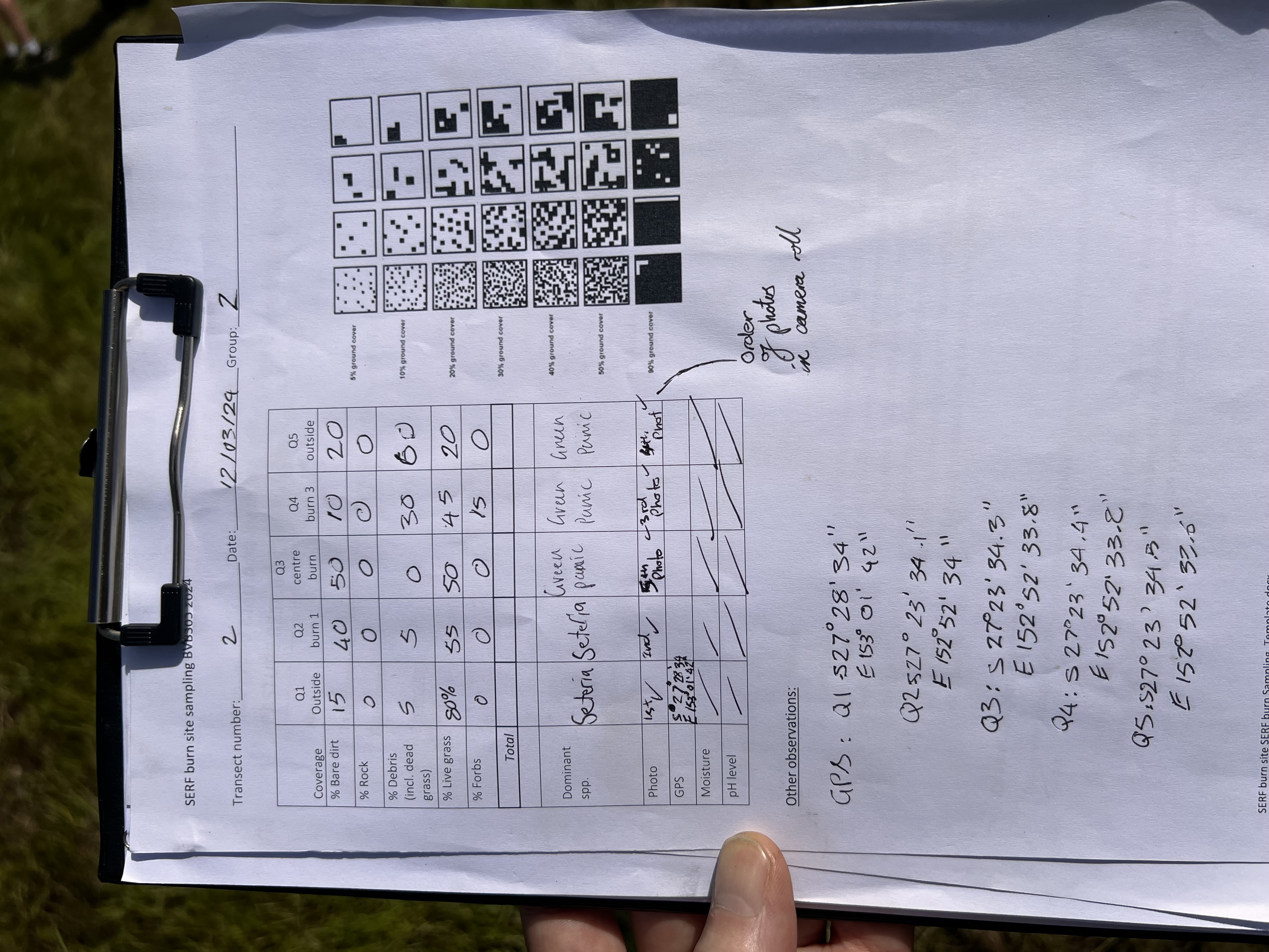
As per the prior study, samples would be taken in transects across the whole site (5 points per transect spanning the strip that we burned last year). Her students and postgraduate team had been analysing those soil samples for the presence of Purpureocillium (see prior soil biology investigation post).
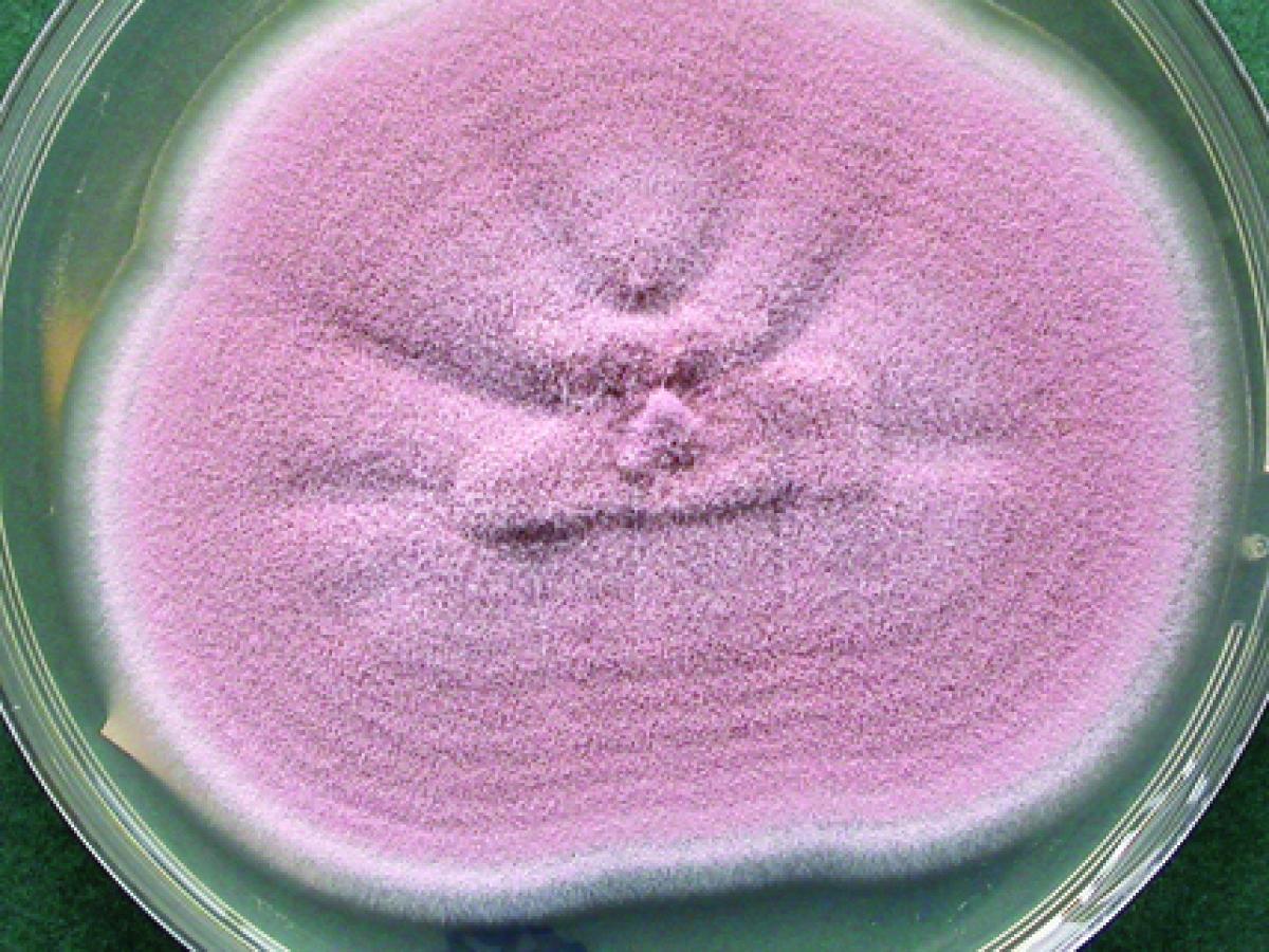
Purpureocillium lilacinum is commonly isolated from soil, decaying vegetation, insects, nematodes and as a laboratory contaminant. It is also a causative agent of infection in human and other vertebrates (Luangsa-ard et al. 2011).

During our discussions we learnt that she would be continuing this work into 20924 – and she invited Eleanor and I to join her class which we have now done on a few occasions. The intentions of her study were to pursue three themes ..
- Soil Ecology: The isolation of facultative root endophytes, particularly P. lilacinum, to determine the effects of burning on abundance and diversity, and diversity within P. lilacinum.
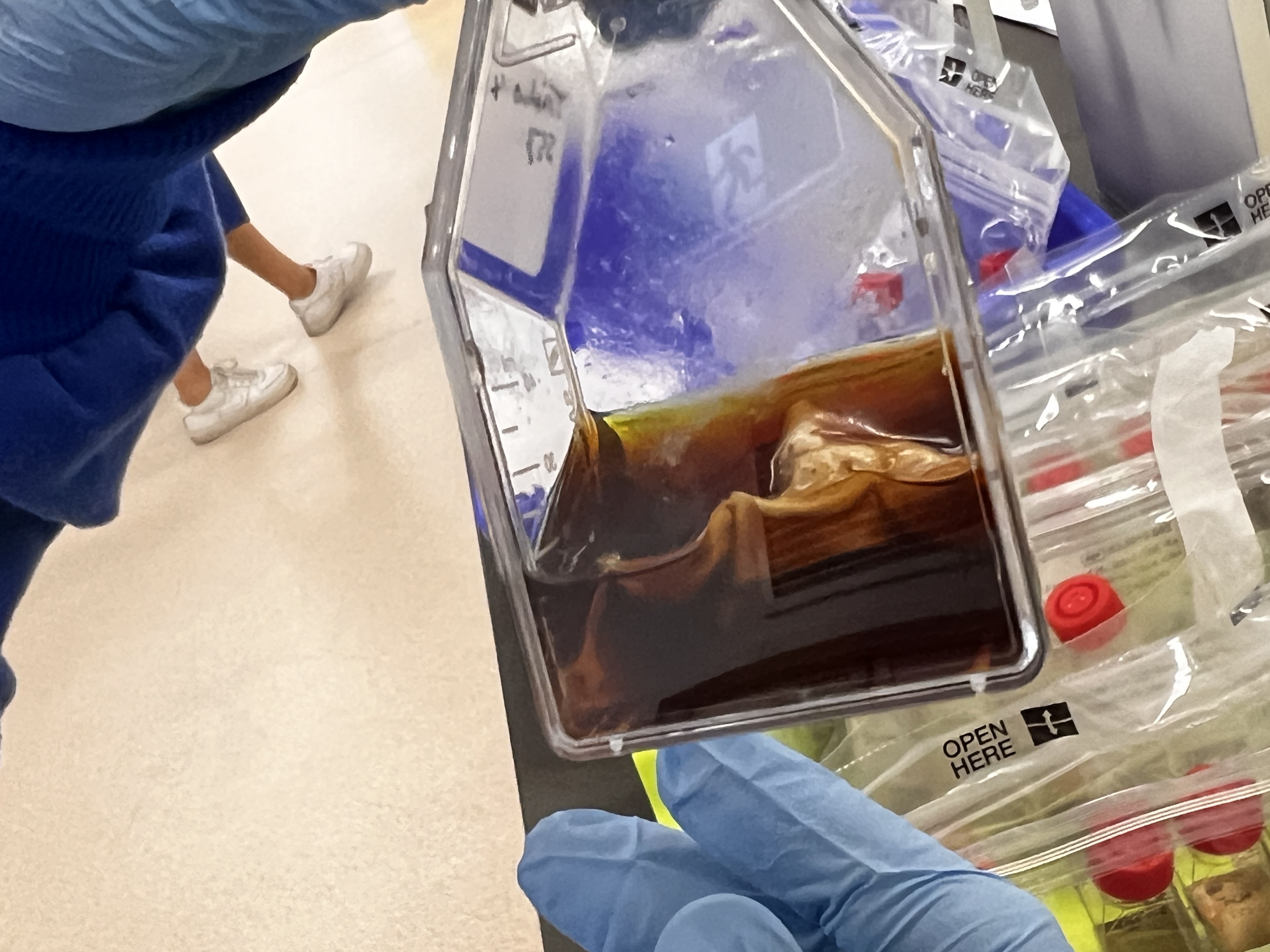
- Media Development: Development of media for the production of P. lilacinum variants as an inoculum (that is – a liquid solution of the fungi that roots can grow through to make them resistant to Mealy bug) – which leads to the third area of interest
- The Good Bugs: The application of P. lilacinum variants as an inoculant against pasture mealybug.
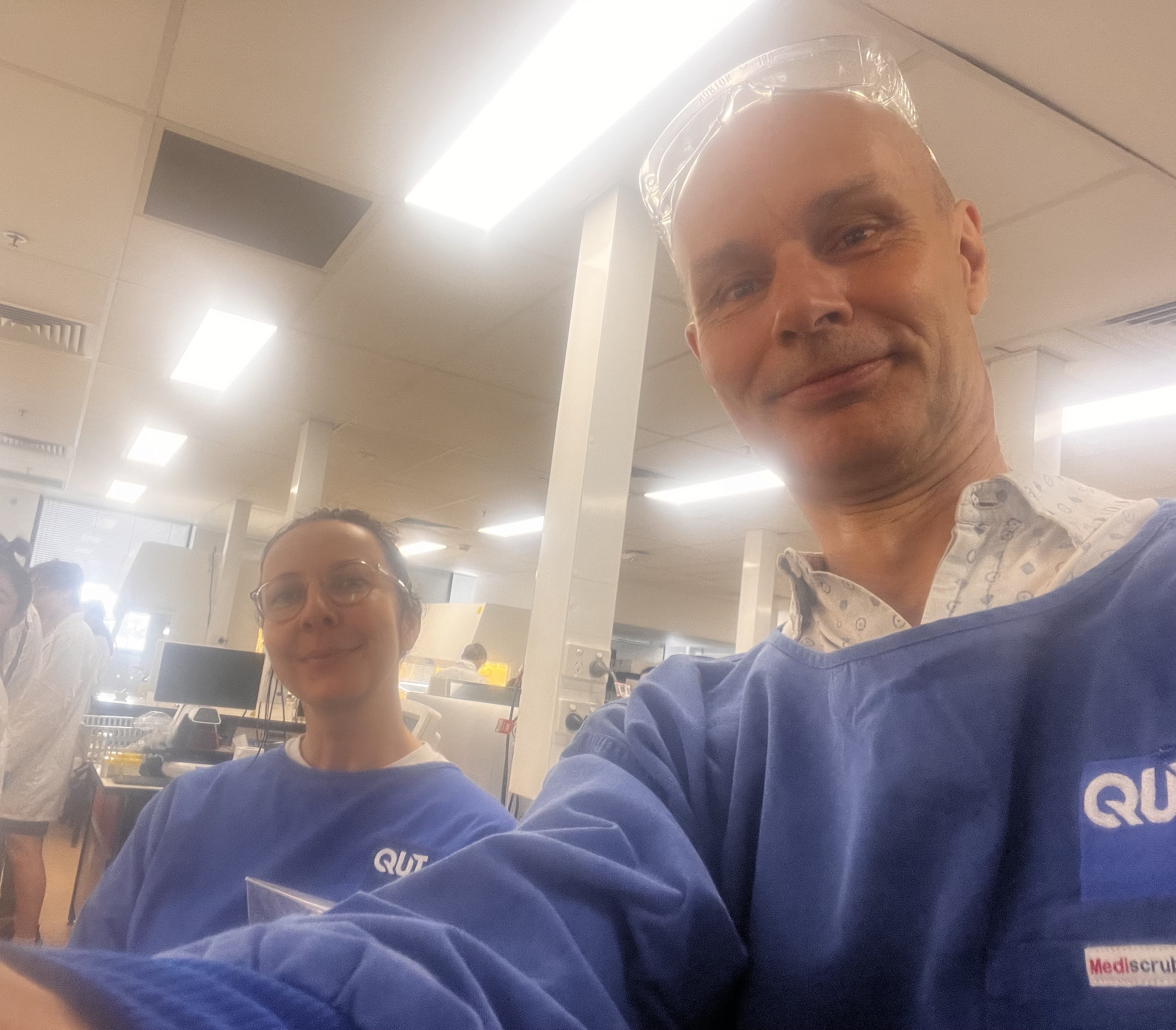
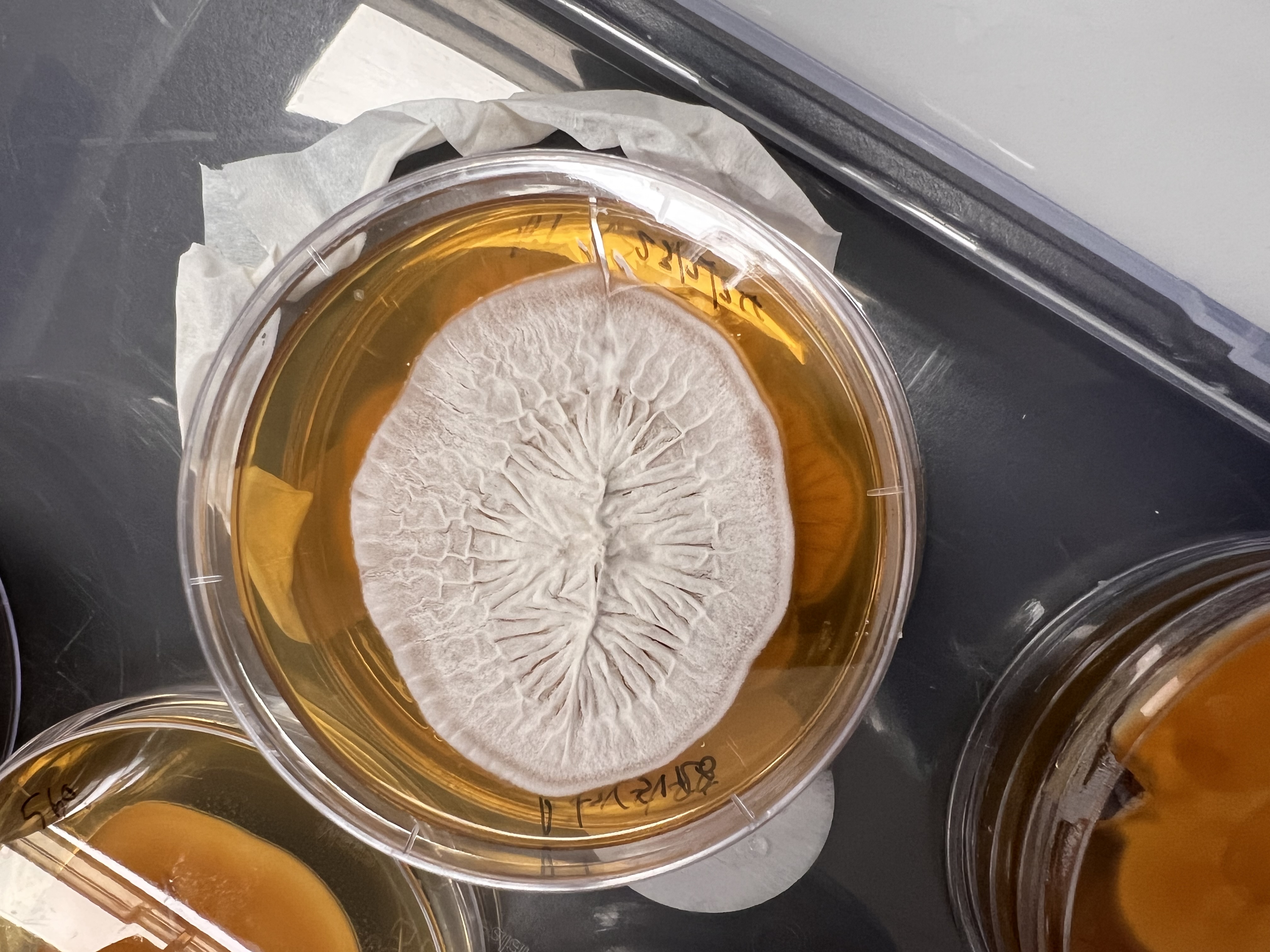
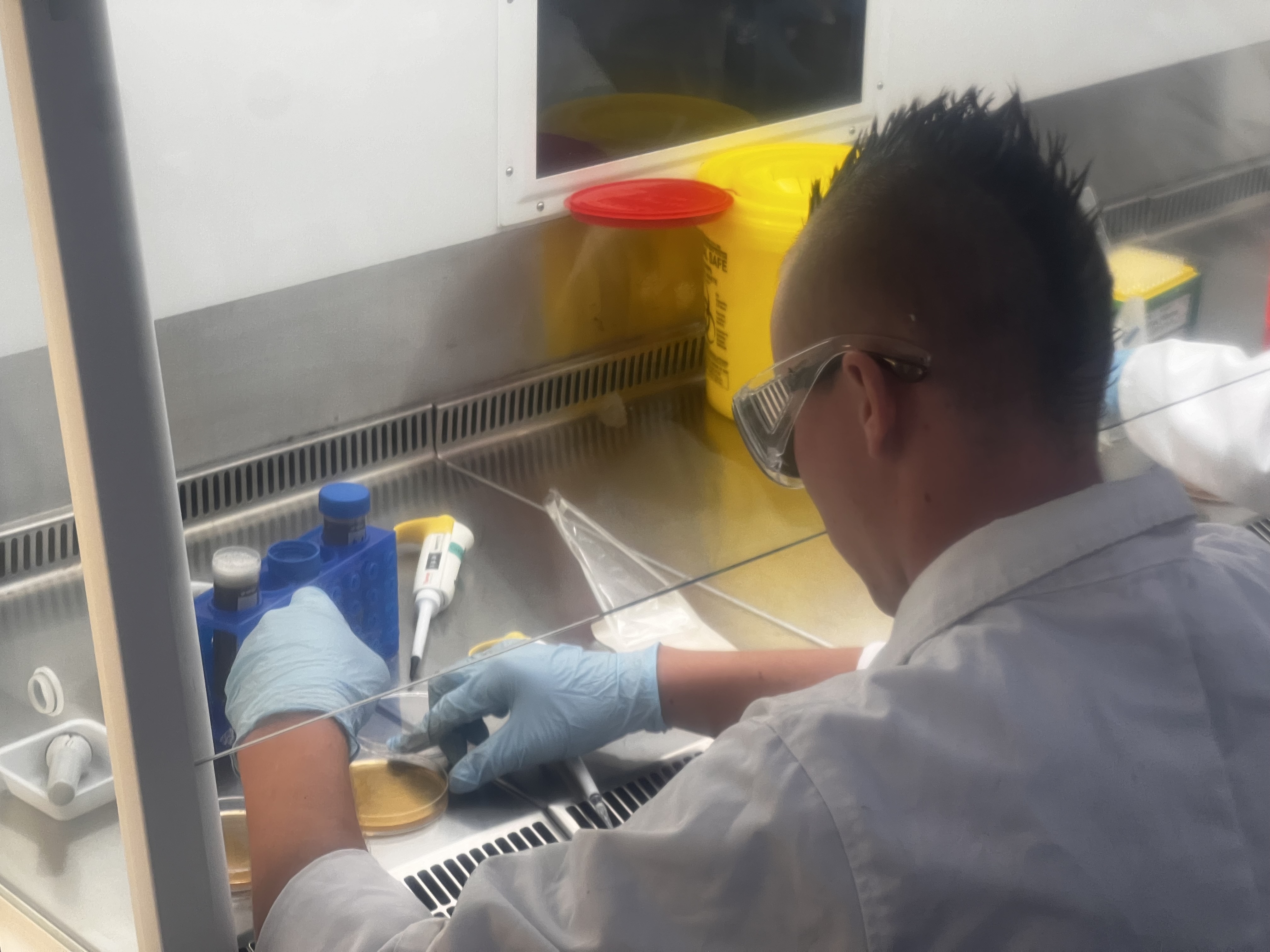
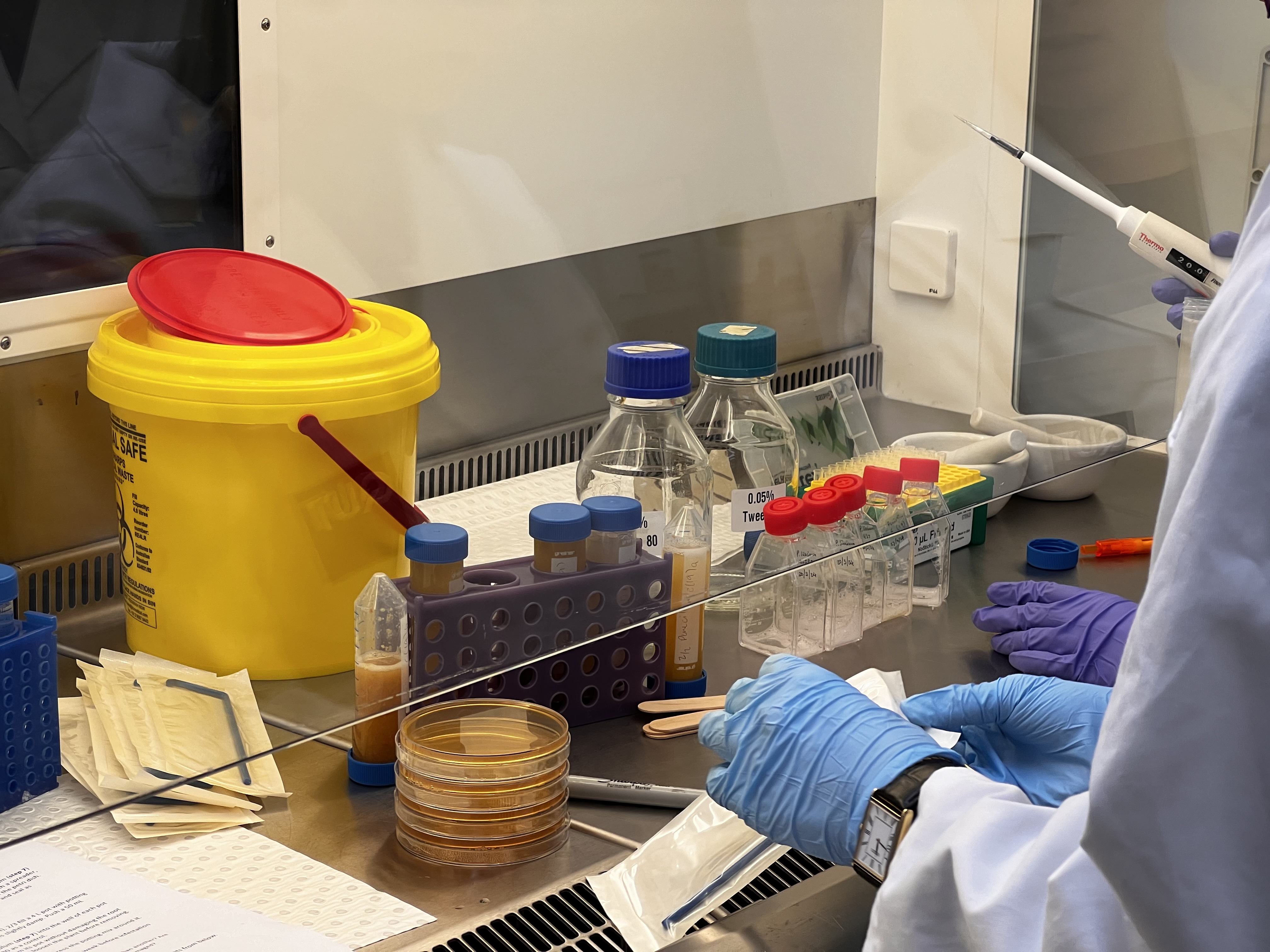
For me it has been a fascinating return to the analogue chemistry methods I remember only from Year 10 (!) – and has included both sampling soil to the required protocol at SERF and then observing its analysis, culturing and the isolation of so called ‘morphotypes’ (in essence any of a group of different types of individuals of the same species in a population/individual fungi) for further growth on agar plate cultures. (An agar plate is a petri dish that contains a growth medium solidified with agar, and is used to culture microorganisms).
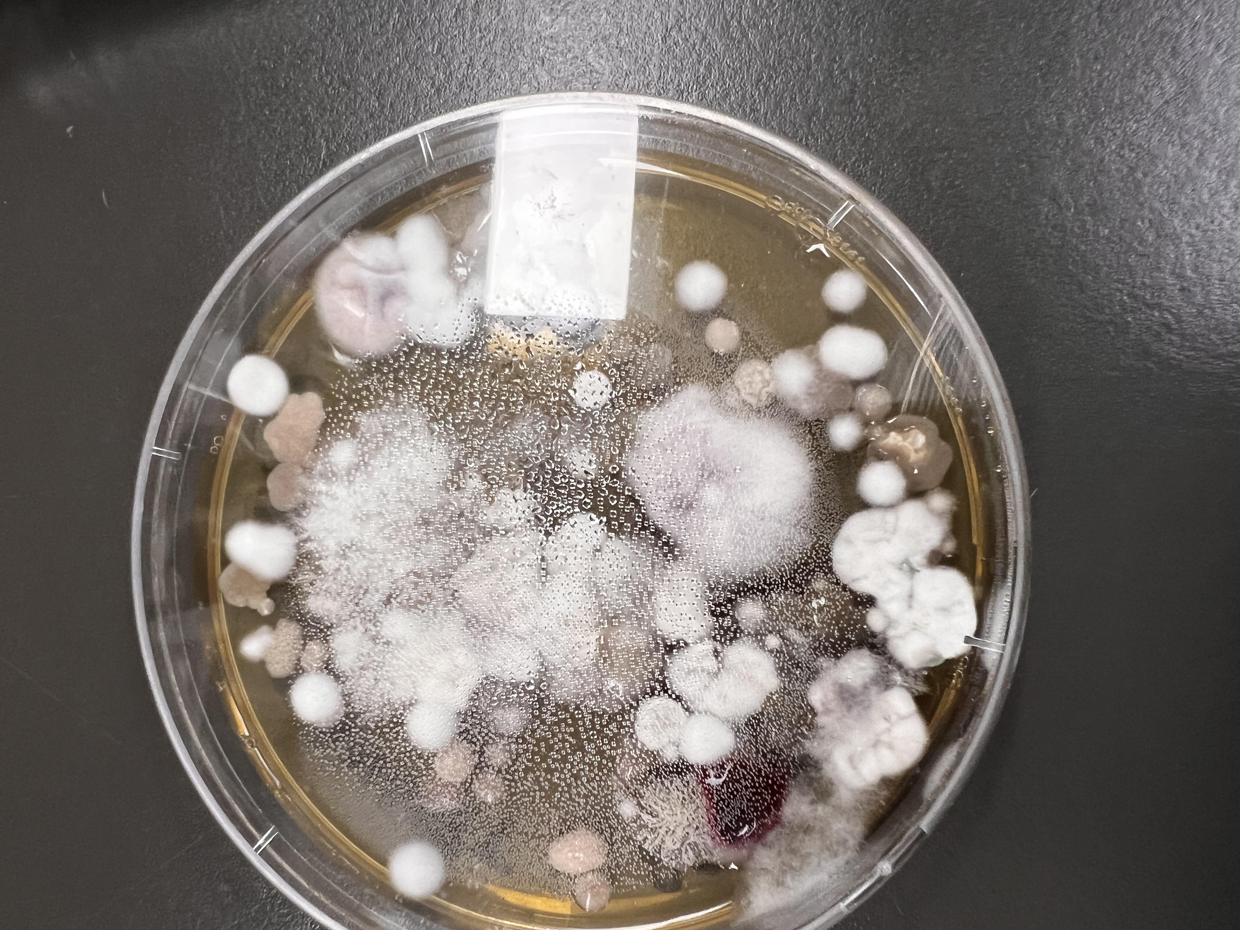
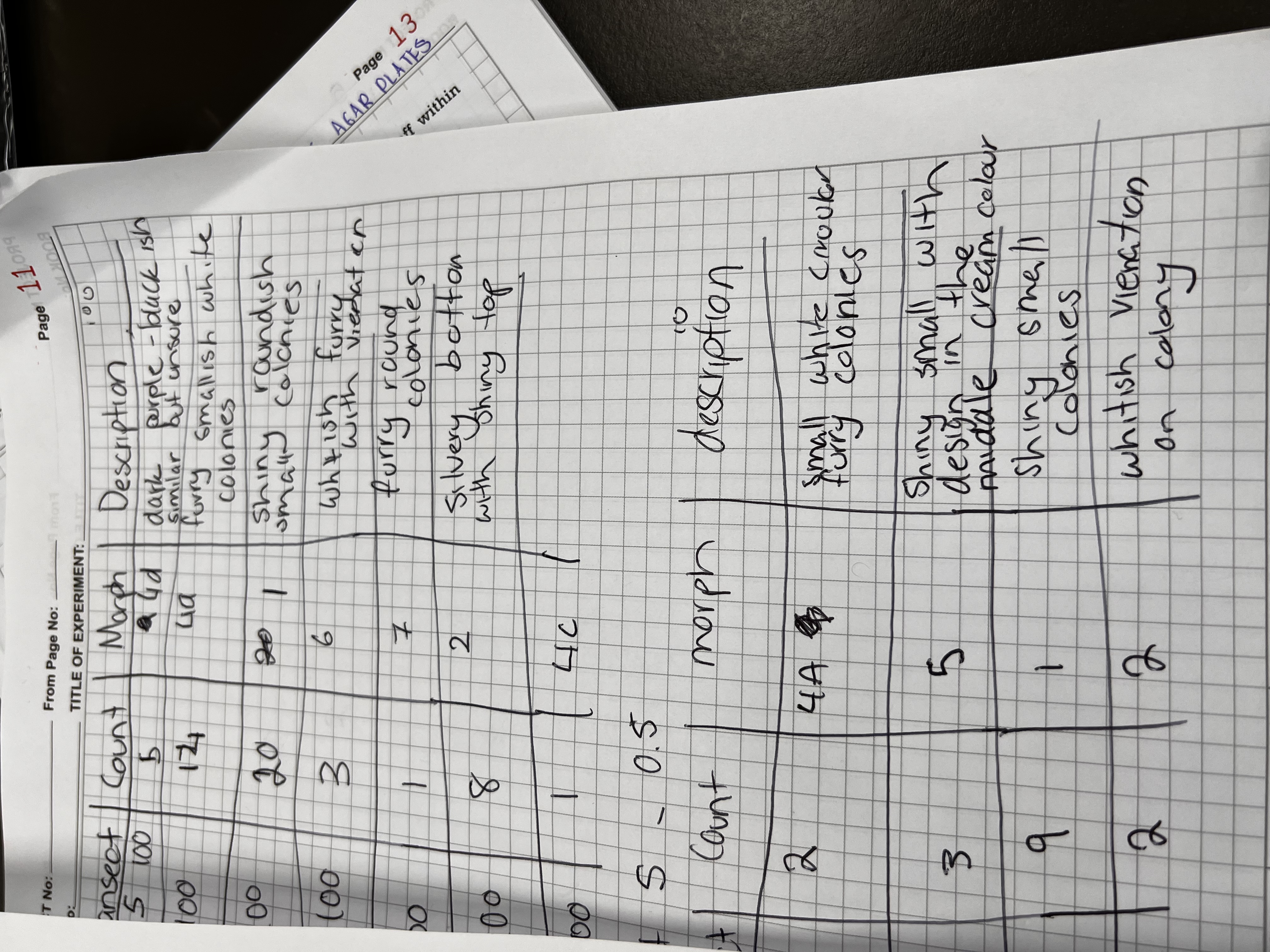
The descriptions alone are evocative and the structure, form and ‘intelligence’ of this form suggest fruitful investigation ahead!
The exciting idea that Caroline has raised – which further cements the fruitful connections emerging between the arts and sciences, is “to do a longitudinal study of the soil microbiome by adapting the sampling this year (across the burn site) to compare the dryer slope with the burned gully below.
Her (and our) interest would therefore be to see the changes in soil fungal diversity over time, particularly once trees are established.” At this stage she is looking to do this highly technical, lab based work over several years – which will allow this project an extraordinary look into the health of the soil as the artwork develops. Exciting times ahead – so many thanks to A/Prof Hauxwell for your kindness, interest and engagement on this co-beneficial process 🙂